Back Of Skull Anatomy / MBBS Medicine (Humanity First): Skull Anatomy / The ethmoid bone forms the central part of the floor, which is the deepest area of the anterior cranial fossa.
Back Of Skull Anatomy / MBBS Medicine (Humanity First): Skull Anatomy / The ethmoid bone forms the central part of the floor, which is the deepest area of the anterior cranial fossa.. Foramina inside the body of humans and other animals. From an anatomical perspective, the skull is divided into two parts: Skull anatomy divides this patchwork of bones into two categories: The foramen magnum, housing the brainstem, is also a part of the. The ethmoid bone forms the central part of the floor, which is the deepest area of the anterior cranial fossa.
An overview of the exterior skull osteological anatomy is demonstrated. The cranium and the mandible. It offers protection to the brain, eye balls, inner ears, and nasal passages. This view of the skull is dominat. The foramen magnum, housing the brainstem, is also a part of the.
The Human Skull Anatomical Chart - Anatomy Models and ... from www.shopanatomical.com The skull is a skeletal framework of the head of vertebrates, that supports the face and makes a protective cavity concerning the brain. The skull bones can be classified into two groups: The ethmoid bone forms the central part of the floor, which is the deepest area of the anterior cranial fossa. Understanding the human skull anatomy is necessary for a wide range of professionals from doctors (dentists, oral surgeons, neurosurgeons, etc.) to the structure of the skull bones is to a large extent determined by and interconnected with the anatomy of the sensory organs, situated in the head, as. The foramen magnum, housing the brainstem, is also a part of the. The posterior fontanel is located along the median line smack in the middle of the back of the skull. The skull includes the upper jaw and the cranium. They don't move and united into a single unit.
The skull begins to form prior to week 12 of embryogenesis.
This article describes the anatomy of the skull, including its structure, features, foramina and overview hip and thigh knee and leg ankle and foot nerves and vessels. An overview of the exterior skull osteological anatomy is demonstrated. This view of the skull is dominat. Human skull from the front. The skull has a single occipital condyle.7 the skull consists of five major bones: This anatomic region is complex and poses surgical challenges for otolaryngologists and neurosurgeons alike. The skull bones can be classified into two groups: The separation of the cranial bone plates at time of birth facilitate passage of the head of the fetus through the mothers birth canal or p. From an anatomical perspective, the skull is divided into two parts: The cranium and the mandible. Understanding the human skull anatomy is necessary for a wide range of professionals from doctors (dentists, oral surgeons, neurosurgeons, etc.) to the structure of the skull bones is to a large extent determined by and interconnected with the anatomy of the sensory organs, situated in the head, as. It supports and protects the face and the brain. Skull, skeletal framework of the head of vertebrates, composed of bones or cartilage, which form a unit that protects the brain and some sense organs.
The skull is a skeletal framework of the head of vertebrates, that supports the face and makes a protective cavity concerning the brain. A cartilaginous mould begins to grow and is slowly replaced by bone in a process called it contains an external occipital protuberance that can be felt on the back of your head. The frontal, parietal, temporal and occipital bones are joined at the cranial sutures. Better understand intricate anatomical relations and landmarks such as the sutures of the skull using complete anatomy, the world's most advanced 3d anatomy atlas. Skull bones aren't fused together at birth.
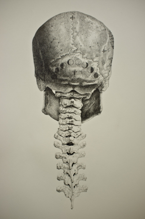
spine-tattoo | Tumblr from 40.media.tumblr.com The frontal (top of head), parietal (back of head), premaxillary and nasal (top beak), and. The skull or known as the cranium in the medical world is a bone structure of the head. They don't move and united into a single unit. Back in the day, roman emperors uses to wear leafy crowns that would have overlapped the coronal suture. The skull is a bony structure that supports the face and forms a protective cavity for the brain. The skull has a single occipital condyle.7 the skull consists of five major bones: Foramina inside the body of humans and other animals. Please feel free to download and print.
Overview, anterior skull base, middle skull base march 18, 2017.
Skull bones aren't fused together at birth. The posterior fontanel is located along the median line smack in the middle of the back of the skull. The skull is a bony structure that supports the face and forms a protective cavity for the brain. Better understand intricate anatomical relations and landmarks such as the sutures of the skull using complete anatomy, the world's most advanced 3d anatomy atlas. This view of the skull is dominat. The cranium and the mandible. Radiographic atlas of skull and brain anatomy. It supports and protects the face and the brain. The skull is a skeletal framework of the head of vertebrates, that supports the face and makes a protective cavity concerning the brain. It offers protection to the brain, eye balls, inner ears, and nasal passages. The skull performs vital functions. They don't move and united into a single unit. The skull base is the inferior portion of the neurocranium.
Radiographic atlas of skull and brain anatomy. The skull performs vital functions. This is a model of the human (homo sapiens) skull. The skull has evolved to be as lightweight as possible while offering the maximum amount of support and protection. It offers protection to the brain, eye balls, inner ears, and nasal passages.
Jonathan Rush - Human Skull Study from cdnb.artstation.com The skull has a single occipital condyle.7 the skull consists of five major bones: This anatomic region is complex and poses surgical challenges for otolaryngologists and neurosurgeons alike. Skull anatomy divides this patchwork of bones into two categories: The ethmoid bone forms the central part of the floor, which is the deepest area of the anterior cranial fossa. Overview, anterior skull base, middle skull base march 18, 2017. The skull begins to form prior to week 12 of embryogenesis. The skull or known as the cranium in the medical world is a bone structure of the head. The bbc is not responsible for the content of external websites.
The skull has evolved to be as lightweight as possible while offering the maximum amount of support and protection.
This is a model of the human (homo sapiens) skull. Skull anatomy divides this patchwork of bones into two categories: The frontal (top of head), parietal (back of head), premaxillary and nasal (top beak), and. The foramen magnum, housing the brainstem, is also a part of the. Understanding the human skull anatomy is necessary for a wide range of professionals from doctors (dentists, oral surgeons, neurosurgeons, etc.) to the structure of the skull bones is to a large extent determined by and interconnected with the anatomy of the sensory organs, situated in the head, as. Skull, skeletal framework of the head of vertebrates, composed of bones or cartilage, which form a unit that protects the brain and some sense organs. The cranium and mandible was exported from ct data. It was then cleaned, adapted and polypainted this model is part of a comparison with the skull of a human. Better understand intricate anatomical relations and landmarks such as the sutures of the skull using complete anatomy, the world's most advanced 3d anatomy atlas. The ethmoid bone forms the central part of the floor, which is the deepest area of the anterior cranial fossa. Overview, anterior skull base, middle skull base march 18, 2017. The skull or known as the cranium in the medical world is a bone structure of the head. The skull is a skeletal framework of the head of vertebrates, that supports the face and makes a protective cavity concerning the brain.


0 Komentar